How to Get Rid of Earring Keloids: A Clinical & Material Guide
Learn how to get rid of earring keloids with clinical treatments, implant-grade jewelry swaps, and expert aftercare to prevent recurrence.

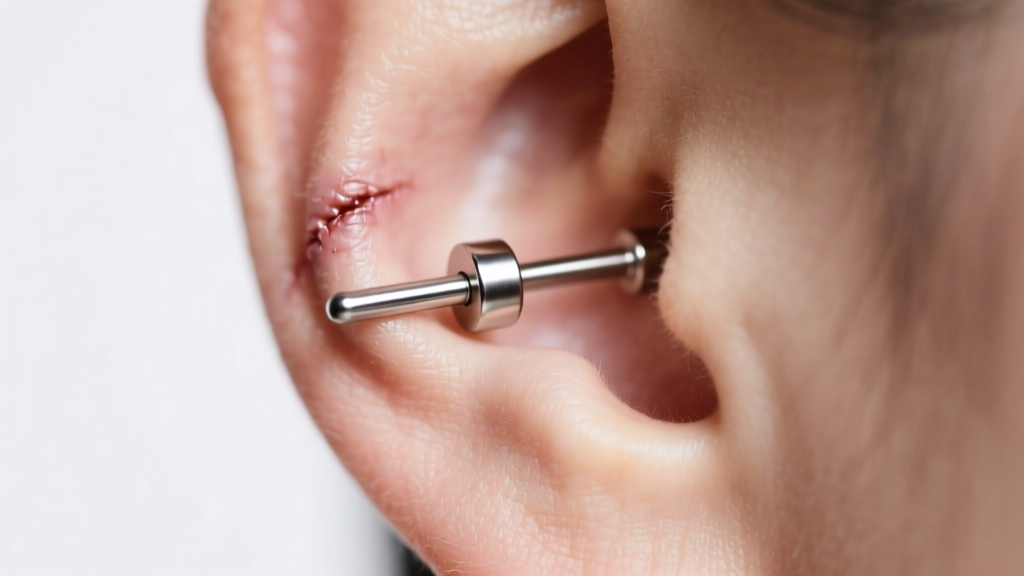
The Pathophysiology of Piercing Keloids vs. Hypertrophic Scars
Understanding how to get rid of earring keloids begins with accurate identification. True keloids are benign fibroproliferative tumors that extend beyond the original boundaries of the piercing trauma. They are driven by an abnormal wound-healing cascade where fibroblasts overproduce Type I and Type III collagen, failing to undergo normal apoptosis. According to the National Institutes of Health (StatPearls), keloids occur in up to 16% of individuals with Fitzpatrick skin types IV through VI, compared to less than 1% in lighter skin types.
Conversely, hypertrophic scars remain confined to the piercing site and often regress spontaneously over 12 to 18 months. Hypertrophic bumps are frequently caused by mechanical irritation, improper jewelry angles, or contact dermatitis. Misdiagnosing a hypertrophic irritation bump as a keloid often leads to unnecessary surgical interventions. A clinical dermatological evaluation is critical before attempting invasive removal, as the surgical excision of a true keloid without adjuvant therapy carries a recurrence rate of up to 80%.
Clinical Interventions: Medical Approaches to Keloid Reduction
When addressing true keloid tissue, over-the-counter remedies are entirely ineffective. You must rely on targeted medical interventions that disrupt the collagen synthesis cycle. Dermatologists and plastic surgeons utilize a tiered approach depending on the lesion's volume and vascularity.
Intralesional Corticosteroid Injections (ILK)
The first-line treatment for small-to-medium earlobe keloids is the injection of Triamcinolone acetonide (Kenalog), typically at concentrations of 10 to 40 mg/mL. The corticosteroid inhibits fibroblast proliferation and increases collagen degradation. Injections are administered every 3 to 4 weeks using a 30-gauge needle. Patients usually require 3 to 6 sessions to achieve significant flattening. A common side effect is localized hypopigmentation and subcutaneous fat atrophy, which is why precise intralesional placement is mandatory.
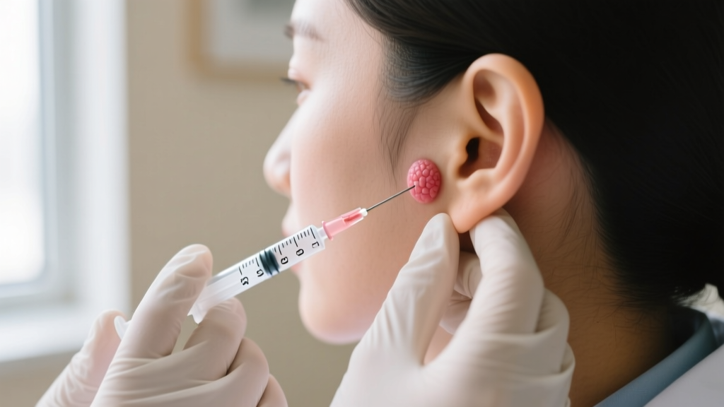
Cryotherapy and Surgical Excision
For bulky, fibrotic keloids that do not respond to ILK, intralesional cryotherapy using a specialized probe freezes the tissue from the inside out, inducing necrosis while sparing the overlying epidermis. Surgical excision is reserved for massive lesions but must never be performed as a standalone procedure. The American Academy of Dermatology (AAD) strongly recommends combining surgical removal with post-operative corticosteroid injections or superficial radiation therapy (SRT) to suppress the inevitable inflammatory rebound.
| Treatment Modality | Mechanism of Action | Recurrence Rate | Est. Cost Per Session (USD) |
|---|---|---|---|
| Intralesional Corticosteroids | Inhibits fibroblast DNA synthesis | 50% - 70% (Monotherapy) | $150 - $300 |
| Intralesional Cryotherapy | Cellular necrosis via freezing | 30% - 40% | $250 - $500 |
| Surgical Excision + SRT | Physical removal + radiation | 10% - 20% | $1,500 - $3,500 |
| 5-Fluorouracil (5-FU) | Antimetabolite blocking cell division | 40% - 50% | $200 - $400 |
The Metallurgical Factor: Upgrading to Implant-Grade Jewelry
A massive percentage of what patients believe are early-stage keloids are actually severe allergic contact dermatitis reactions leading to hypergranulation. The culprit is almost always low-grade "surgical steel." The term surgical steel is unregulated in the jewelry industry and often encompasses 316L stainless steel, which contains up to 14% nickel. When exposed to the saline environment of a healing fistula, nickel ions leach into the tissue, triggering a localized immune response.
To eliminate this variable, you must transition to ASTM F136 Implant-Grade Titanium (Ti 6AL-4V ELI) or pure, unalloyed Niobium. These materials are entirely nickel-free and biocompatible. Furthermore, the gauge of the jewelry plays a critical mechanical role. Standard 20-gauge or 18-gauge earrings act like a wire cheese-cutter on compromised tissue, creating continuous micro-tears that stimulate collagen production. Upgrading to a 16-gauge or 14-gauge internally threaded or threadless labret stud distributes the physical load across a wider surface area, drastically reducing mechanical tension on the fistula.
Pressure Therapy and Mechanotransduction
Once a keloid has been flattened via corticosteroid injections or excision, maintaining the result requires physical intervention. Pressure therapy operates on the principle of mechanotransduction. By applying continuous, localized pressure (typically 20-30 mmHg) to the scar bed, you induce local hypoxia. This hypoxia downregulates the expression of Transforming Growth Factor-beta (TGF-β), the primary cytokine responsible for keloid formation.
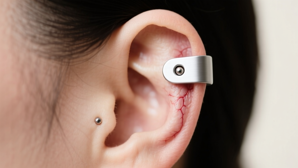
Specialized magnetic pressure earrings or custom-molded silicone Zimmer splints are worn for 12 to 24 hours a day for a minimum of 6 to 12 months post-treatment. While cumbersome, pressure therapy reduces the recurrence rate of surgically excised keloids by nearly half when used consistently. Silicone gel sheets can also be applied to the posterior and anterior lobes to maintain hydration and regulate epidermal temperature, further signaling the fibroblasts to cease excessive collagen production.
Post-Treatment Aftercare & Recurrence Prevention
After successfully reducing a keloid, the micro-environment of the piercing must be strictly managed to prevent a rebound. Avoid all essential oils, including tea tree oil, which is a known sensitizer and can trigger contact dermatitis in compromised tissue. The only approved topical cleanser is sterile, isotonic 0.9% sodium chloride wound wash, applied once daily.
Sleep positioning is another hidden variable in recurrence. Side-sleepers place continuous compressive and shearing forces on the earlobe, which can aggravate the healing dermal matrix. Utilizing a travel pillow or a specialized piercing pillow with a central cavity allows the ear to remain suspended, eliminating nocturnal friction. Finally, avoid changing the jewelry prematurely. The fistula of a previously keloid-prone ear remains highly volatile for up to 18 months; leaving the initial implant-grade titanium labret in place undisturbed is the most effective long-term prevention strategy.
"The successful eradication of an earring keloid is never achieved through a single modality. It requires a triad approach: precise dermatological intervention, uncompromising metallurgical standards, and relentless mechanical aftercare."


