How To Cure An Earring Infection: A Clinical Piercing Guide
Learn how to cure an earring infection with clinical precision. Expert advice on saline ratios, titanium grades, and identifying rejection vs. infection.

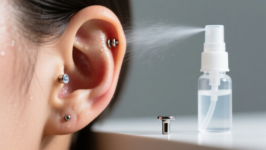
When a fresh or healed piercing becomes inflamed, panicked internet searches often lead to outdated and potentially harmful home remedies. Understanding how to cure an earring infection requires a clinical approach that differentiates between true bacterial pathogens, mechanical irritation, and metallurgical allergies. As of 2026, professional piercers and dermatologists emphasize evidence-based aftercare protocols over harsh chemical treatments. This guide provides a deep dive into the pathophysiology of piercing infections, exact triage frameworks, and the material science required to restore tissue health.
The Pathophysiology of Piercing Infections
A piercing is essentially a puncture wound that creates a fistula (a tube of scar tissue). When the skin barrier is compromised, opportunistic bacteria can colonize the tract. The two most common culprits in earring infections are Staphylococcus aureus and Pseudomonas aeruginosa.
- Staphylococcus aureus: A gram-positive bacterium naturally residing on the skin. It typically causes localized, warm, and painful lobe infections with thick, yellowish exudate.
- Pseudomonas aeruginosa: A gram-negative bacterium that thrives in moist environments. It is notoriously common in cartilage piercings and is characterized by a sweet, grape-like odor and a distinct greenish-blue purulent discharge.
Treating the infection correctly hinges on identifying the bacterial profile and addressing the environmental factors—such as jewelry friction or moisture trapping—that allowed the colony to proliferate.
Triage: Infection vs. Irritation vs. Rejection
Before attempting to cure an earring infection, you must confirm that an infection is actually present. According to the Association of Professional Piercers (APP), up to 80% of suspected infections are actually mechanical irritation bumps or allergic contact dermatitis. Use the clinical triage table below to assess your symptoms.
| Condition | Primary Trigger | Exudate Profile | Pain & Sensation | Clinical Intervention |
|---|---|---|---|---|
| Bacterial Infection | Pathogen colonization (Staph/Pseudomonas) | Thick, opaque yellow/green; foul odor | Throbbing, hot to touch, radiating pain | Saline irrigation, topical/oral antibiotics |
| Mechanical Irritation | Snagging, sleeping on it, long posts | Clear, sticky, or pale white (lymph fluid) | Tender, localized sharp pain on contact | Downsize jewelry, warm compresses, rest |
| Allergic Dermatitis | Nickel leaching from low-grade metals | Clear, weeping, crusty, flaky skin | Intense itching, burning, localized rash | Upgrade to ASTM F-136 titanium immediately |
| Migration / Rejection | Immune system treating jewelry as a splinter | None, or minimal clear lymph | Painless or mild tightness; skin thinning | Remove jewelry to prevent severe scarring |
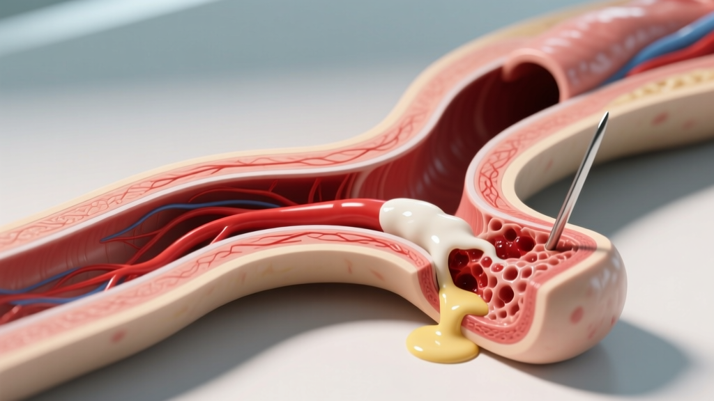
Step-by-Step Clinical Protocol: How to Cure an Earring Infection
If your symptoms align with a true bacterial infection, follow this targeted protocol to eradicate the pathogen while preserving the fistula tract.
1. Isotonic Saline Irrigation (0.9% NaCl)
Flush the piercing twice daily with a sterile 0.9% isotonic saline solution. This specific concentration matches the body's natural osmolarity, preventing cellular dehydration while mechanically washing away bacterial biofilms and necrotic tissue. You can purchase sterile wound wash, or compound it at home using 1/4 teaspoon of non-iodized fine-grain sea salt dissolved in 8 ounces of sterile distilled water. Never use table salt, which contains iodine and anti-caking agents that cause chemical burns.
2. Thermal Compresses for Vascular Dilation
After irrigating, apply a clean, non-woven gauze pad soaked in warm saline for 3 to 5 minutes. The heat induces vasodilation, increasing localized blood flow and delivering white blood cells to the infected cartilage or lobe tissue to fight the pathogen.
3. Post Downsizing and Snag Elimination
Standard earring posts are often 8mm to 10mm long to accommodate initial swelling. Once the acute swelling subsides (usually within 2-3 weeks), the excess post acts as a lever. Every time the earring is bumped, it torques inside the fistula, creating micro-tears that invite secondary infections. Visit a professional piercer to downsize to a snug 5mm or 6mm flat-back labret.
The Role of Jewelry Metallurgy in Healing
You cannot cure an earring infection if the jewelry itself is the source of the trauma. Many commercial earrings are made from 316L "surgical steel," which contains 8% to 12% nickel. In the presence of saline and body heat, nickel ions leach into the tissue, triggering allergic contact dermatitis—a condition that perfectly mimics a bacterial infection. 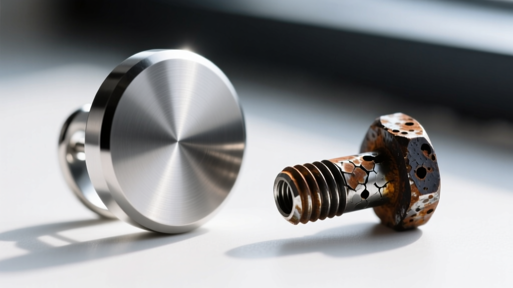
For any compromised piercing, the APP Jewelry Guidelines mandate the use of ASTM F-136 Implant-Grade Titanium or Niobium. These materials are entirely nickel-free, highly polished to a mirror finish (preventing bacterial adhesion), and biologically inert. Upgrading your jewelry is often the single most effective step in resolving chronic piercing "infections" that are actually metal allergies.
Cartilage vs. Lobe: Divergent Healing Timelines and Risks
The anatomical location of your earring dictates the severity and treatment pathway of the infection.
- Earlobe Infections: Lobes are highly vascularized, meaning they have a robust blood supply that delivers immune cells and systemic antibiotics efficiently. Lobe infections generally respond well to rigorous topical saline care and, if necessary, a short course of topical mupirocin or oral antibiotics.
- Cartilage Infections (Perichondritis): Cartilage is avascular; it relies on the perichondrium (the connective tissue covering it) for nutrients. If bacteria breach the cartilage, the resulting infection (chondritis) can rapidly lead to tissue necrosis, abscess formation, and permanent structural deformity (cauliflower ear). Cartilage infections almost always require systemic oral antibiotics, specifically fluoroquinolones like Ciprofloxacin, which target Pseudomonas.
When to Escalate: Systemic Antibiotics and Medical Intervention
While localized redness and minor swelling are expected, certain red flags indicate that the infection has breached the local tissue and is spreading systemically. Seek immediate medical attention from a physician or dermatologist if you experience:
- Expanding Erythema: Redness that spreads outward from the piercing site, especially if accompanied by red streaks (lymphangitis).
- Systemic Symptoms: Fever, chills, nausea, or swollen lymph nodes in the neck and jawline.
- Abscess Formation: A localized, fluctuant pocket of pus that requires professional incision and drainage.
Critical Warning: If you suspect a severe infection, do not remove the earring without consulting a doctor first. Removing the jewelry allows the surface of the fistula to heal over, trapping the bacterial infection inside the tissue and forming a closed abscess that requires surgical intervention. The jewelry acts as a drain; keep it in place until a medical professional advises otherwise.
By combining precise metallurgical choices, strict osmotic saline irrigation, and an understanding of tissue vascularity, you can effectively manage and cure an earring infection while preserving the integrity of your piercing.


